The Promise of PEEK
PEEK is a relative newcomer to medicine, but in some applications, it is already changing how medical professionals treat their patients. This phenomenon is most pronounced in neck and back treatments, where PEEK devices can offer excellent outcomes in many instances.
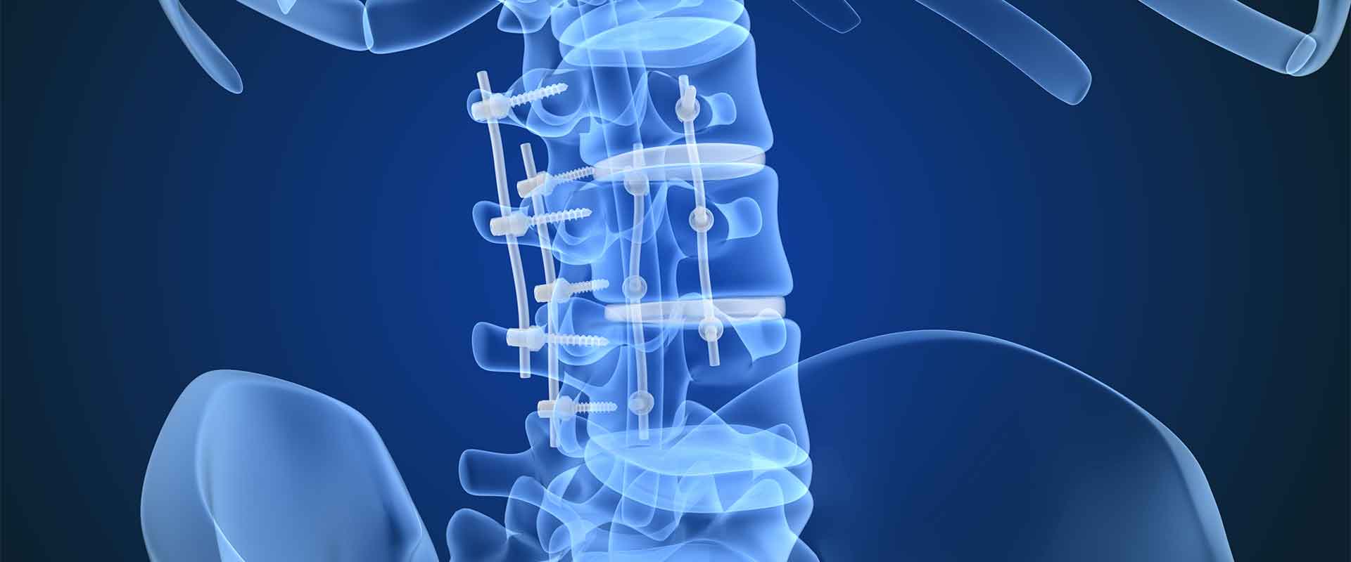
The most common use of PEEK in spinal surgery is in spinal fusions. PEEK spinal cages represent the standard of care in intervertebral and lumbar fusion. PEEK spinal cages have been studied extensively since the early 2000s, and in 2016, they were used in approximately 75 percent of all spinal fusion surgeries. As such, they have quickly replaced titanium and stainless steel devices in this role. There are several reasons for this, and as PEEK continues to assert itself as the most promising biomaterial going forward, it will become the material of choice in many more applications.
PEEK’s Many Uses in Spinal Surgery
The debate between titanium and PEEK in spinal implants is still ongoing, and it’s likely that there won’t ever be a clear winner. Titanium and PEEK bring different qualities to treatment, so the optimal choice depends on the procedure, surgeon and patient.
What makes PEEK such a natural fit for spinal implant and surgical applications? PEEK possesses unique properties that other materials, including titanium, do not offer. Some of those properties include:
1. A similar modulus to bone – PEEK can closely mimic the stiffness of bone, which has a couple of major implications. For one, because it performs like bone, it is easier for medical professionals to predict how the device will work following implantation. As post-operative monitoring is a critical component of spinal surgery outcomes, this added predictability is extremely valuable.
Further, with a similar modulus to bone, PEEK spinal components largely avoid stress shielding. As such, patients with PEEK spinal implants can better maintain bone density in the area around the surgical site.
2. Added flexibility, when needed – PEEK’s bone-like modulus is a key reason it is a valuable addition to the spinal surgery market. In spinal fusion, this modulus stabilizes the vertebrae while preserving enough flexibility for reduced stress on the bone. This dynamic is the reason PEEK is also an emerging option for interspinous spacers and in disc arthroplasty.
In both interspinous spacers and disc arthroplasty, the implant must be flexible enough to facilitate natural movement of the spine. If the implant does not do so, additional stress will be placed on neighboring vertebrae, potentially resulting in stress fractures and other concerning complications.
Although PEEK does not stabilize joints to the degree that titanium can, it does offer an ideal mix of flexibility and arthrodesis. This particular combination makes PEEK an intriguing option for pedicle-based rod systems, as an alternative or adjunct to spinal fusion. This form of treatment can stabilize the spine dynamically, which is only possible because of PEEK’s superior flexibility.
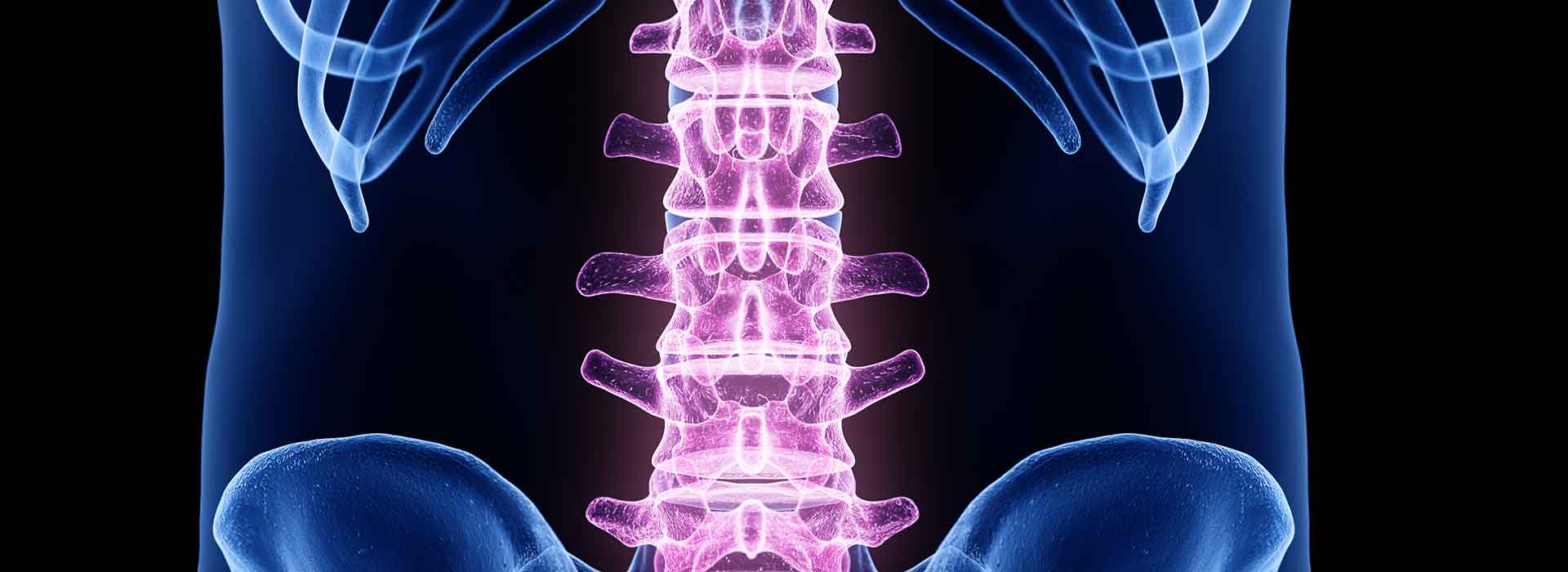
3. Transparency during imaging – Spinal surgeries being as delicate as they are, patients must be frequently monitored to determine treatment efficacy. If there are any complications associated with the implant, they must be detected early on. This degree of vigilance means the patient must be imaged regularly. Imaging, though, is difficult to do with metal implants, as they create high opacity artifacts.
PEEK components are completely radiolucent, or transparent, during imaging, which allows medical professionals to make accurate estimations of the patient’s progress including bone formation and growth.
4. A promising future – PEEK is constantly being scrutinized and researched, and this research is heavily focused on improving bone apposition with PEEK components, as this would further enhance the long-term reliability of PEEK spinal implants.
There are several promising leads to this end, including the addition of hydroxyapatite into the PEEK matrix. Hydroxyapatite is known throughout medicine as an effective osteoconductive material, in that it encourages bone on-growth wherever it is located. PEEK designs with hydroxyapatite are already available in some treatments, and the early returns are positive. Accelerated bone formation and greater amounts of bone formation are both expected in patients who receive this enhanced PEEK implant model.
Additional avenues are being explored to increase this osteoconductive effect including PEEK tissue scaffolds and PEEK additives similar to hydroxyapatites. Another option in encouraging bone on-growth is the use of porous implants. With porous cavities available for bone growth, with time the implant and bone lock together, enhancing implant stability. This can be used in concert with hydroxyapatite-enhanced PEEK implants, improving osteoconductivity further.
Research is also investigating PEEK implants that are designed with antimicrobial in the drive to reduce infections at surgical sites.
5. Fabrication advantages – PEEK can be machined into extremely complex designs at extremely tight tolerances. It can be scaled up or down as needed, so it can be fabricated into components that would pose a challenge to metal component manufacturers.
PEEK can also be injection molded, which allows for mass production of identical, complex device components. With larger production runs and extensive machining capabilities available, medical researchers and professionals can do more with PEEK.
PEEK implantology got its start in spinal devices. Unsurprisingly, PEEK is thoroughly represented in spinal surgical treatments, and its potential applications are only multiplying as material engineering improves on the PEEK front.